- Follow Us
CDI Labs Services for
Target Deconvolution and Target Discovery
HuProt can help guides the development of novel therapies.
REQUEST INFOHuProt for Identifying Specific Molecular Targets
Understand the mechanism of action of the compound and its' effects on biological pathways
Target deconvolution is a process in drug discovery and development that involves identifying the specific molecular target(s) of a monoclonal or polyclonal antibody, bioactive compound or drug candidate. This process is crucial for understanding the mechanism of action of the compound, elucidating its effects on biological pathways, and assessing its potential therapeutic applications.
In disease research, it is not uncommon to identify antibodies without initially knowing their specific target. Monoclonal antibodies with unknown targets may have therapeutic potential for treating diseases or conditions for which the target antigen is unknown or poorly characterized. Further investigation and target identification efforts may reveal novel therapeutic opportunities based on the antibody's binding properties and biological effects.
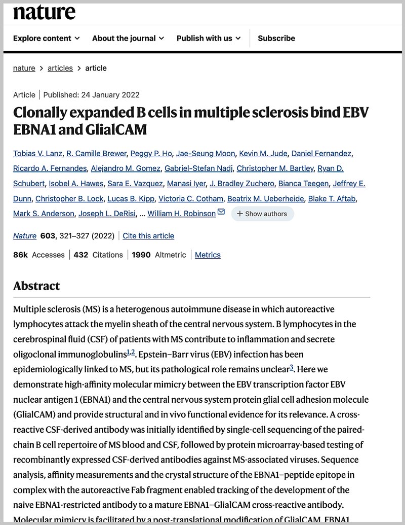
In the featured publication below, HuProt™ microarray and VirScan® PhIP-Seq were used in the featured publication which demonstrated high-affinity molecular mimicry between the EBV transcription factor EBV nuclear antigen 1 (EBNA1) and the central nervous system protein glial cell adhesion molecule (GlialCAM) and provided structural and in vivo functional evidence for its relevance. The results provide a mechanistic link for the association between MS and EBV and could guide the development of new MS therapies.
Clonally expanded B cells in multiple sclerosis bind EBV EBNA1 and GlialCAM
Abstract
Multiple sclerosis (MS) is a heterogenous autoimmune disease in which autoreactive lymphocytes attack the myelin sheath of the central nervous system. B lymphocytes in the cerebrospinal fluid (CSF) of patients with MS contribute to inflammation and secrete oligoclonal immunoglobulins. Epstein–Barr virus (EBV) infection has been epidemiologically linked to MS, but its pathological role remains unclear. Here we demonstrate high-affinity molecular mimicry between the EBV transcription factor EBV nuclear antigen 1 (EBNA1) and the central nervous system protein glial cell adhesion molecule (GlialCAM) and provide structural and in vivo functional evidence for its relevance.
VIEW PAPERCDI Labs Advantages
HuProt Sample Requirements
Human serum or plasma
One 20 μL per sample
Antibody isolates
10 μg per sample (concentration 0.1 mg/mL)
Human cerebrospinal fluid (CSF)
Minimum 1.5 mL per sample
Protein / peptide / small molecule
10 μg per sample (concentration 0.1 mg/mL)
RNA
1 mL per sample (concentration 2 μM)
Other fluids
> Please contact usPhiP-Seq Sample Requirements
Serum or plasma
20 μL aliquot per sample
Cerebrospinal fluid (CSF)
250 μL aliquot per sample
Other antibodies (IgG monoclonals, B cell supernatants, etc)
250 μL aliquot per sample (0.01 mg/mL concentration)
12 sample minimum
In multiples of x12 samples (i.e., x12, x24, x36, 48x – full rows of a standard 96-well plate), and any number after 48 samples

